

For Brands & Sellers
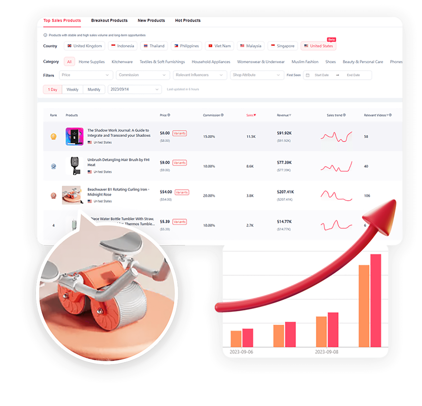
Find & track winning products
Shoplus help you find niche with hourly-updated data of product sales on TikTok, you can find products with maximum profit potential and craft your product strategy in more effecient way.
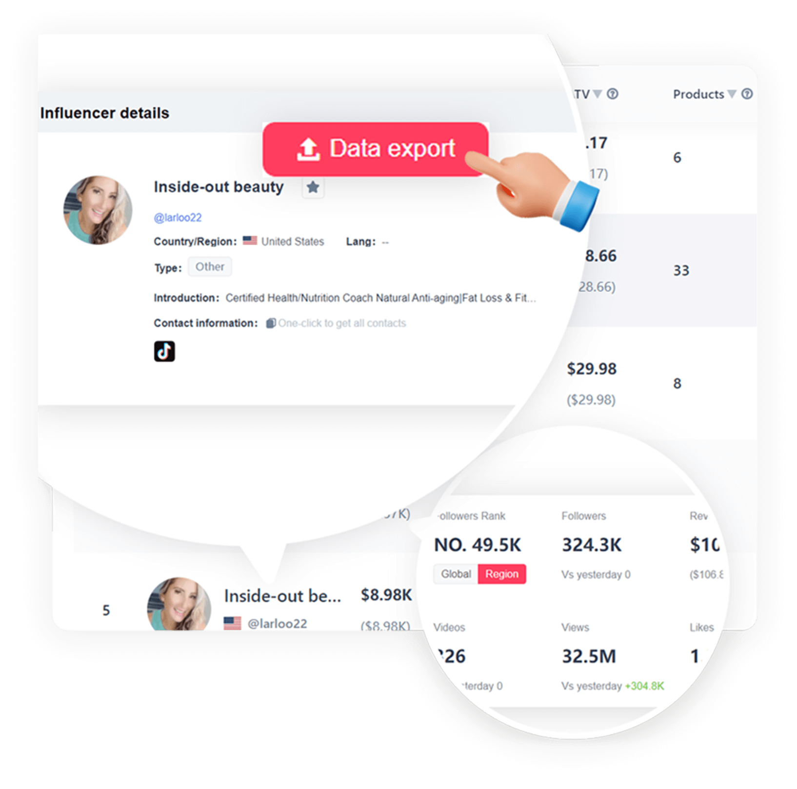
Make conscious decisions for influencer marketing
Better understanding of TikTok influencer marketing performance with data-driven insight. Save, monitor and compare all engagement and sales metrics of top TikTok influncers side by side.
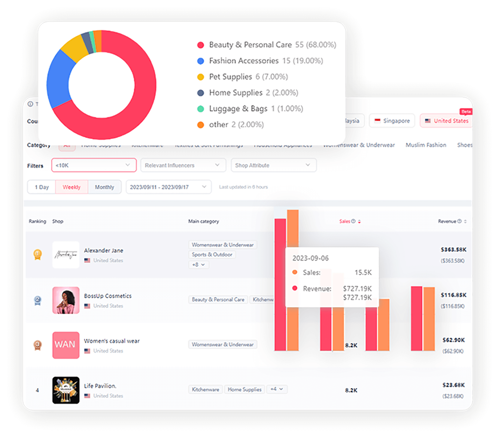
Spy on competitors with TikTok data analysis
Gain a competitive and accurate data analysis of TikTok Shop. Knowing your rivals' product, sales, and marketing strategies to optimize your own business strategies.
- Get StartedView Plans


Find & track winning products
Shoplus help you find niche with hourly-updated data of product sales on TikTok, you can find products with maximum profit potential and craft your product strategy in more effecient way.
Make conscious decisions for influencer marketing
Better understanding of TikTok influencer marketing performance with data-driven insight. Save, monitor and compare all engagement and sales metrics of top TikTok influncers side by side.
Spy on competitors with TikTok data analysis
Gain a competitive and accurate data analysis of TikTok Shop. Knowing your rivals' product, sales, and marketing strategies to optimize your own business strategies.
- Get StartedView Plans


For TikTok Creators
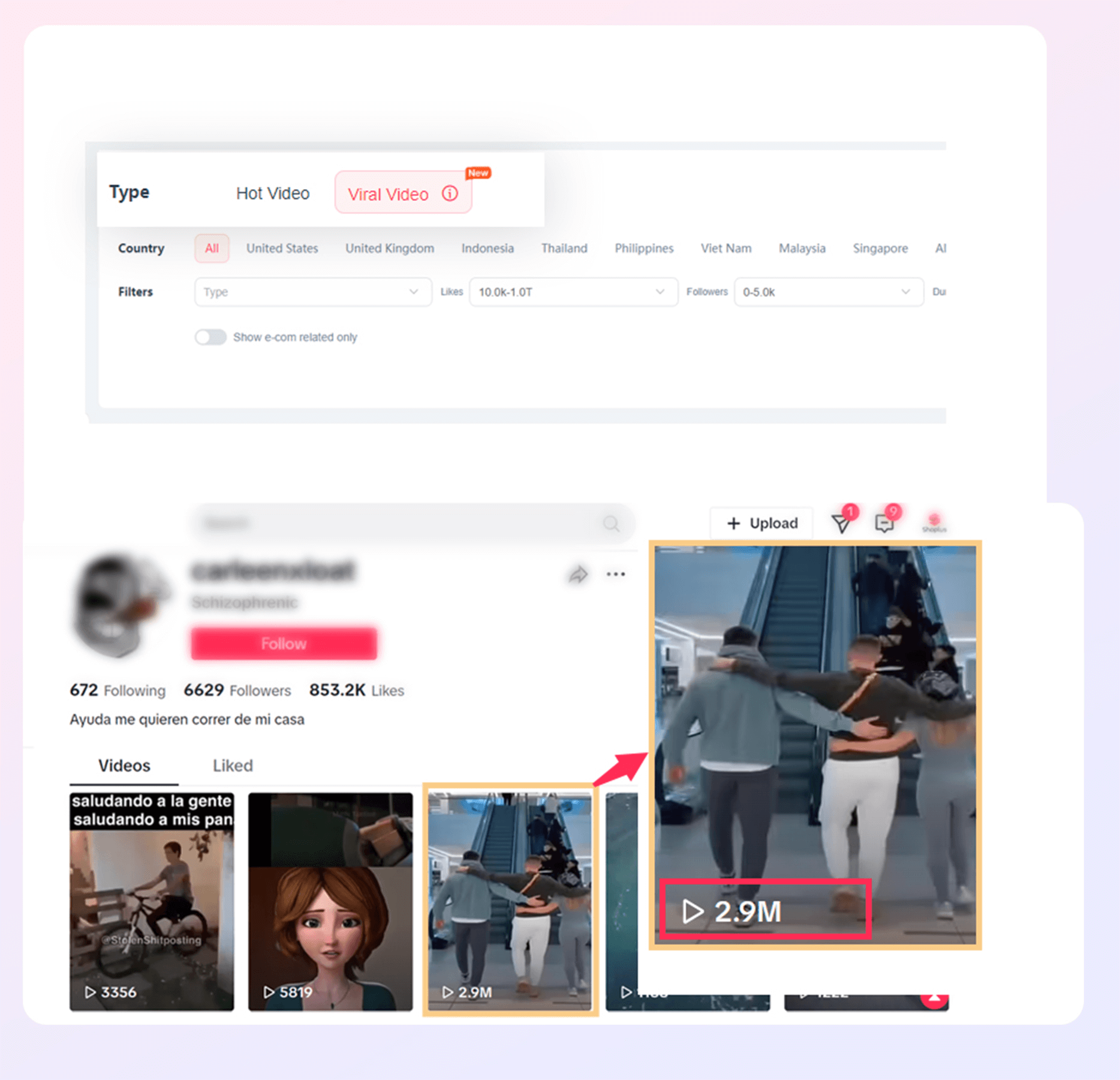
Discover viral and hot video
Struggling with views under 1,000? Dive into the "Viral Video" feature and emulate the success of your peers. It’s time for you to get TikTok famous. We've identified over 10,000 renowned creators and trending videos in your niche for you.
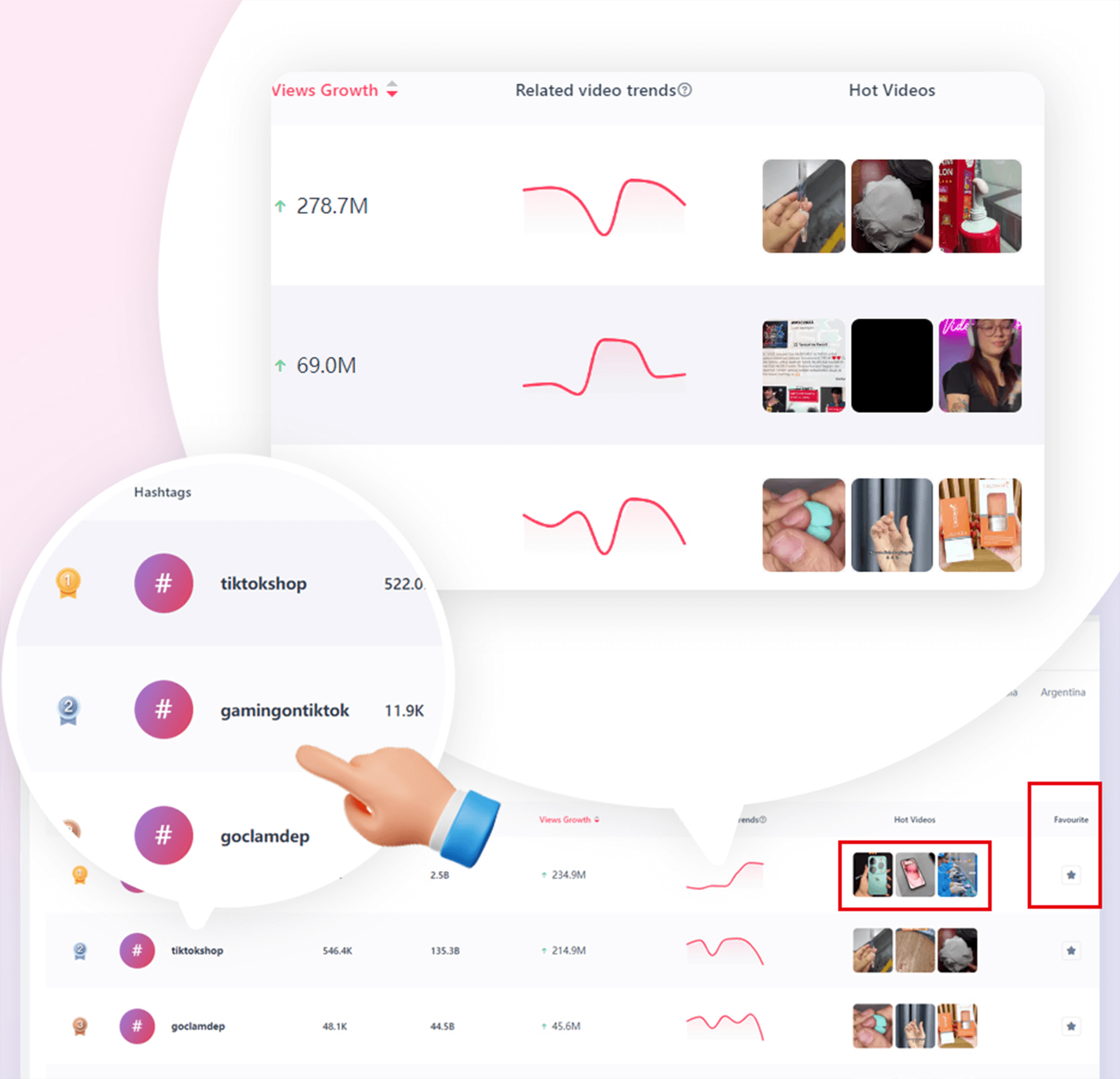
Hashtags list with trending insights
Explore the latest TikTok trends with hashtags.We offer trending hashtag insights to help you grow your followers.
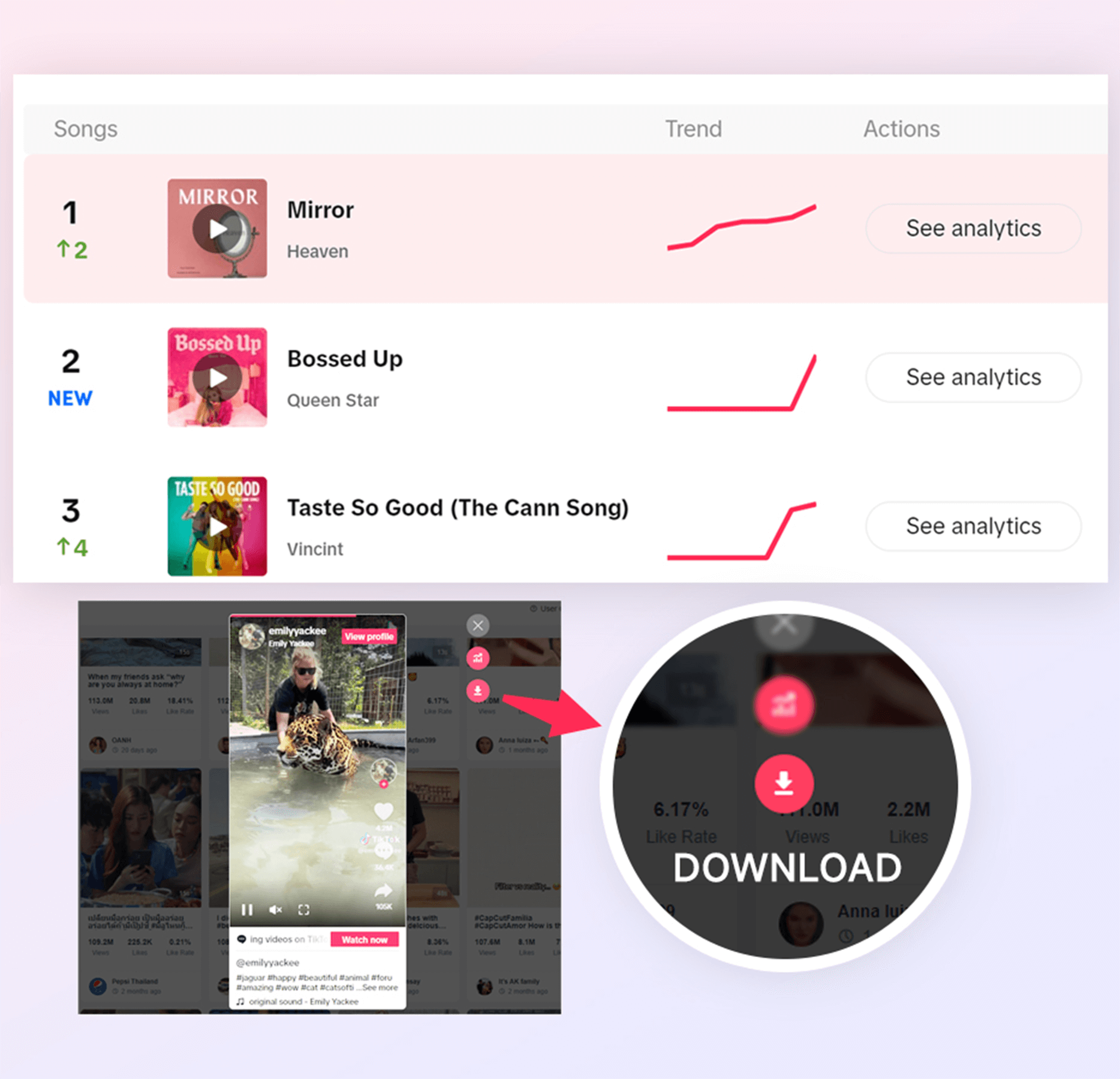
Save viral videos, music, and hashtags in just one click
Tired of hunting for trending sounds or mimicking videos when creating content? Save your favorite sounds and clips easily, and seamlessly craft your next viral video.
- Get StartedView Plans


Discover viral and hot video
Struggling with views under 1,000? Dive into the "Viral Video" feature and emulate the success of your peers. It’s time for you to get TikTok famous. We've identified over 10,000 renowned creators and trending videos in your niche for you.
Hashtags list with trending insights
Explore the latest TikTok trends with hashtags.We offer trending hashtag insights to help you grow your followers.
Save viral videos, music, and hashtags in just one click
Tired of hunting for trending sounds or mimicking videos when creating content? Save your favorite sounds and clips easily, and seamlessly craft your next viral video.
- Get StartedView Plans